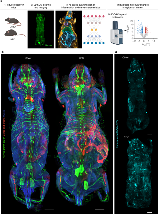
This study introduces a breakthrough deep-learning framework called MouseMapper, designed to analyze how diseases such as obesity affect the entire body at cellular resolution. MouseMapper quantitatively analyzes neural networks, immune cells, and 31 organs and tissues, making it possible to understand systemic disease-driven changes with much higher precision. The framework revealed changes in neural structure and immune-cell distribution in obese mice, and it suggests that obesity-related sensory dysfunction and inflammatory responses may appear similarly in humans. This technology could open new possibilities for studying complex diseases and developing therapies.
1. Why Whole-Body Pathology Analysis Is Needed, and Why Existing Tools Fall Short
Lifestyle-related diseases such as obesity are known to affect many organ systems throughout the body. These systemic effects show how interconnected human physiology is and why it is important to understand pathological changes holistically. Until now, however, researchers lacked tools that could study whole-body perturbations at the cellular and molecular level, making it difficult to grasp the breadth of those effects.
Recent tissue-clearing methods combined with fluorescence microscopy can visualize whole mice or large human tissue samples at single-cell resolution. That is impressive, but it creates a new problem: there has not been a good way to analyze and quantify the enormous image datasets that result. Elongated structures such as nerves, tissues, and organs are especially hard to analyze at whole-body scale. Existing image-analysis methods are often limited to particular organs or become unstable when imaging resolution and labeling strategies change. A new tool was needed to overcome these limitations and reveal the mechanisms behind systemic disease effects.
2. The Birth of MouseMapper, an Innovative Deep-Learning Framework
To meet that need, the research team developed MouseMapper. The framework segments and analyzes whole-body images of the nervous and immune systems using three-dimensional image analysis. It also helps select regions of interest for downstream molecular analysis. MouseMapper consists of three major modules.
- Nerve-Module: Quantitatively analyzes and maps peripheral neural networks.
- Immune-Module: Segments immune cells and quantifies their distribution.
- Tissue-Module: Maps segmented structures to 31 organs and tissues so quantitative data can be compared and interpreted biologically.
One of MouseMapper's biggest strengths is that it generalizes robustly across different imaging resolutions and antibody-labeling datasets without retraining. In other words, it can analyze images accurately even when they were produced under different conditions. Using MouseMapper, the researchers successfully identified structural changes in nerve and immune-cell networks at high spatial resolution. In particular, they found structural changes in the infraorbital branch of the trigeminal nerve in obese mice. These changes were linked to whisker sensory dysfunction and proteomic changes associated with axon degeneration. Remarkably, the same molecular features were preserved in the trigeminal ganglia of obese humans.
2.1. How MouseMapper Was Developed and the Core Technologies Behind It
MouseMapper was built to quantify obesity-induced whole-body changes without bias. The framework analyzes transparent whole-mouse images and 3D reconstructions produced using vDISCO clearing and light-sheet fluorescence microscopy.
-
Nerve module:
- The research team manually annotated nerves in a virtual reality environment to create training data. With that data, they fine-tuned VesselFM, a foundation model specialized for 3D vessel segmentation. Because vessels and nerves share similar morphological characteristics, the model worked effectively for nerve analysis as well.
- The model maintained high segmentation accuracy across different image resolutions and labeling strategies, including antibody labeling. It successfully distinguished elongated axonal structures regardless of signal intensity or labeling method.
-
Immune module:
- The team again used VR to annotate Cd68-eGFP+ cells and build training data.
- The fine-tuned foundation model outperformed other deep-learning networks. It also showed strong generalization, successfully segmenting Cd68-eGFP+ cells in tissues such as liver and intestine that were not included in the initial training set. This means it handled signal heterogeneity and dense cell clusters well.
-
Tissue module:
- For organ mapping, where cell-level accuracy is less critical, the tissue module used image downsampling to improve processing speed and efficiency.
- The researchers used VR to annotate 27 organs across 12 whole-mouse scans, and a 3D UNet architecture performed best.
- For adipose and muscle tissue, downsampled images did not preserve texture information well, so the team trained those separately using full-resolution data.
- The resulting tissue module generates a comprehensive anatomical map of 31 organs and tissues, with far greater coverage and accuracy than other state-of-the-art analysis frameworks.
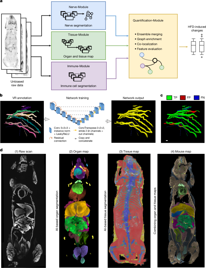
MouseMapper provides a powerful automated AI pipeline for detecting and quantifying system-wide perturbations in neural structures and immune-cell distributions across the whole body.
3. Neural Changes Observed in Obese Mice and Their Functional Impact
Obesity is closely associated with many neurological dysfunctions, especially peripheral neuropathy. But until now, the systemic changes obesity causes in peripheral nerves had not been comprehensively characterized. The team used MouseMapper's nerve and tissue modules to analyze whole-body scans of control and obese mice.
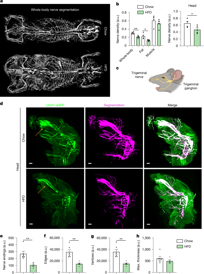
The results showed that obese mice had reduced whole-body nerve density. In adipose tissue, nerve density did not increase in proportion to the increased fat mass; instead, it decreased. This suggests that obesity suppresses innervation.
The most notable finding was a reduction in nerves in the head region. The infraorbital nerve, a branch of the trigeminal nerve that innervates the whisker pad, showed clear structural changes. In mice fed a high-fat diet, the number of nerve endings, edges, and vertices decreased by 60.7%, 57.8%, and 57.6% respectively, while nerve thickness remained similar. This suggests a defect in axonal extension far from the ganglion rather than simple degeneration of the whole nerve.
To understand the functional meaning of these structural changes, the researchers performed a whisker-stimulation test. Obese mice showed a markedly reduced response to whisker stimulation.
"Obese mice showed a markedly reduced response to whisker stimulation."
This strongly suggests that structural changes in facial branches of the trigeminal nerve may contribute to obesity-related sensory dysfunction.
4. Proteomic Changes in the Trigeminal Ganglion: Clarifying Molecular Mechanisms
To uncover the molecular mechanisms behind the infraorbital nerve changes, the researchers performed spatial proteomic profiling of the trigeminal ganglion, where the neuronal cell bodies of the infraorbital nerve are located.
The analysis found 230 proteins that were abnormally regulated in the trigeminal ganglia of mice fed a high-fat diet: 67 upregulated and 163 downregulated.
Pathway analysis showed dysregulation in pathways related to actin cytoskeleton regulation, RHO GTPase effectors, and axon guidance. This may indicate disruption of actin dynamics, which are essential for maintaining axonal structure and function. Pathways related to inflammation and cellular stress responses, including complement and coagulation cascades, ERB signaling, and sphingolipid signaling, were also significantly dysregulated.
One especially important finding was the downregulation of SERPIN-A family proteins, which help protect tissue from inflammation-related damage. This may mean that the ability to control inflammatory tissue damage and the breakdown of structural proteins essential to neural integrity is reduced.
Even more striking, similar molecular changes appeared in postmortem trigeminal ganglion tissue from obese humans. In humans, pathways related to axon guidance, neurodegeneration, and actin cytoskeleton regulation were also dysregulated. This suggests that the MouseMapper findings may translate to human pathology.
"The preservation of these key molecular features in the trigeminal ganglia of obese humans shows the translational value of our study."
These proteomic changes help explain structural changes in the infraorbital nerve, highlight the connection between obesity and neuroinflammation, and may point toward new therapeutic targets.
5. Spatial Understanding of Obesity-Induced Systemic Inflammation
Chronic inflammation is a major feature of obesity and is closely related to the development of many chronic diseases throughout the body. To understand obesity-induced inflammation, it is important to know which tissues and organs are affected and to what degree. The team used MouseMapper's immune and tissue modules to study the spatial context of inflammation in obese mice.
Cd68-eGFP+ immune cells were observed as round clusters in several tissues, including adipose tissue, liver, skeletal muscle, and abdominal wall. The size of immune-cell clusters can indicate inflammatory status inside tissue: larger clusters are associated with more inflammatory and pro-inflammatory states.
The researchers divided Cd68-eGFP+ clusters into three size categories: small clusters with fewer than 6 cells, medium clusters with 6-60 cells, and large clusters with more than 60 cells.
- In obese mice fed a high-fat diet, the proportion of small clusters decreased significantly in liver, visceral fat, and stomach.
- In contrast, the proportion of medium clusters increased in liver and visceral fat, suggesting a shift from small to medium clusters.
- Large clusters increased significantly in several sites, including subcutaneous fat, visceral fat, muscle, stomach, and abdominal wall. This indicates intensified inflammatory activity and immune-cell involvement in obesity.
Multilabel imaging revealed close spatial interactions between T cells and antigen-presenting macrophages, dendritic cells, or B cells within macrophage-rich clusters. Macrophages most frequently colocalized with T cells, NK cells, and endothelial cells, forming perivascular immune hubs.
"Analysis of CD68+ cluster-size changes with MouseMapper confirmed an elevated inflammatory state across tissues in high-fat-diet-induced obesity and provided detailed spatial information."
These results clearly show the pattern of systemic inflammatory responses caused by obesity and make it possible to understand, in spatial detail, the degree and composition of immune-cell accumulation in specific tissues.
6. MouseMapper's Strengths, Limitations, and Future Outlook
6.1. MouseMapper's Major Strengths
MouseMapper provides a powerful and scalable blueprint for comprehensively analyzing perturbations across whole-body systems.
- Comprehensive analysis: It can study disease-induced changes at cellular resolution through 3D organ and tissue mapping, without predefined regions of interest.
- High accuracy: It can accurately segment centimeter-long nerve structures across the body and identify immune cells from single cells to clusters of hundreds of cells.
- Foundation-model approach: The nerve and immune modules fine-tune foundation models pretrained on large 3D biomedical datasets, giving them high segmentation accuracy and generalization across resolutions and antibody-labeling strategies.
- True 3D analysis: Instead of relying on 2D projections or pre-cropped volumes, MouseMapper performs true 3D sliding-window inference on uncropped multi-terabyte datasets, enabling unbiased screening of whole-body anatomical changes.
- Integrated multi-system analysis: Unlike previous methods that focus on a single biological system, MouseMapper integrates peripheral nerves, immune cells, and 31 tissue compartments. That is especially useful for studying complex systemic diseases such as obesity.
"MouseMapper makes it possible to see whole-body changes at the cellular level in a way that was previously impossible."
6.2. Current Limits and Future Development
MouseMapper is a major advance, but whole-body light-sheet microscopy still has limitations.
- Resolution limits: The standard configuration, a 1.1x objective with 0.1 NA, covers the whole body while providing cellular-level detail. But it cannot fully resolve the thinnest parenchymal nerve fibers or subcellular structures such as axons only a few micrometers wide. These small elements appear bundled or merged at the current voxel size, so detailed reconstruction of terminal dendritic branches or synaptic boutons remains beyond whole-body imaging.
- Data volume: Higher-resolution imaging, for example a 4x objective with 0.35 NA, can detect thinner axons and isolated immune cells more accurately. But it can produce up to 50 TB of data per mouse, creating a trade-off between spatial resolution and scalability.
These limits may be overcome through advances in optics, adaptive sampling strategies, and data-processing methods. MouseMapper may also require additional fine-tuning to generalize fully across many imaging modalities.
6.3. Conclusion and Future Uses
MouseMapper identified site-specific neuropathy in obesity, connected it to functional and molecular changes, and demonstrated relevance to human pathology. That is an important turning point in understanding complex disease.
"MouseMapper is a very powerful tool for comprehensively understanding changes across whole-body systems and identifying potential therapeutic targets."
The pipeline can be readily applied to other whole-body systems, such as the lymphatic or vascular systems, and to other complex diseases. Combined with spatial proteomic analysis of structural-change hotspots, MouseMapper can help identify potential therapeutic targets for reversing or preventing pathological change.
The research team made all data and algorithms available online so more scientists can use the technology: https://discotechnologies.org/MouseMapper/. Scientists can now explore enormous datasets from obese and control mice through this online map, quickly examine obesity-induced nerve and immune-cell changes in tissues or organs of interest, and investigate potential connections with other body systems.
MouseMapper provides a powerful blueprint for holistic analysis of complex biological phenomena in 3D and is expected to contribute significantly to future disease research and therapy development.